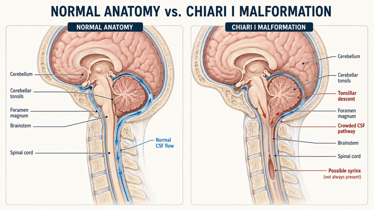
By Jennifer Richards, LMT
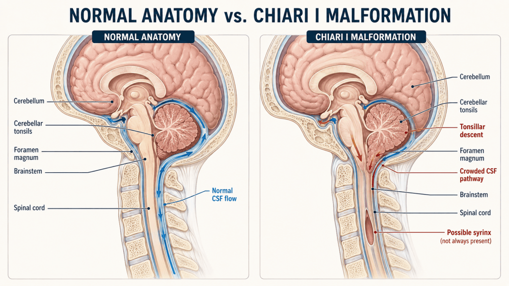
Chiari malformation is sometimes described as “the brain being too large for the skull.” That is an oversimplification, but it captures the basic issue: structures at the back and bottom of the brain are crowded near the opening at the base of the skull.
For massage therapists, the diagnosis does not automatically mean that massage is prohibited. It does mean that treatment must be individualized. The therapist needs to understand the client’s type of Chiari malformation, current symptoms, related neurological conditions, surgical history and medical restrictions before deciding whether—and how—to proceed.
What Is Chiari Malformation?
Chiari malformation is a structural disorder involving the cerebellum, brainstem and foramen magnum. The cerebellum sits at the lower back of the brain and helps coordinate balance, posture and movement. The foramen magnum is the large opening at the base of the skull through which the brainstem continues into the spinal cord.
In Chiari malformation, part of the cerebellum extends lower than expected toward or into the spinal canal. This crowding may compress nearby neurological structures or interfere with the normal circulation of cerebrospinal fluid (CSF), the fluid that surrounds and cushions the brain and spinal cord.
The presence of low-lying cerebellar tonsils on an MRI does not, by itself, prove that Chiari is causing a person’s symptoms. Some people have significant tonsillar descent and few or no symptoms, while others may have clinically important crowding or disrupted CSF flow with a smaller measurement. Symptoms, neurological findings, associated conditions and imaging must be considered together.
The Main Types
● Type I is the most common form encountered in adolescents and adults. The cerebellar tonsils extend below the foramen magnum. It is often present from birth but may not be discovered until later in life. Many cases are found incidentally.
● Type II involves more extensive displacement of the cerebellum and brainstem and is strongly associated with myelomeningocele, a severe form of spina bifida. This is the form historically called Arnold–Chiari malformation.
● Type III is rare and severe. Brain tissue extends through an abnormal opening in the skull or upper spine.
● Type IV refers to severe underdevelopment of the cerebellum and is extremely rare.
Some specialists also use terms such as Chiari 0 or Chiari 1.5, but classification and terminology can vary. For most adult massage clients, Type I is the form most likely to appear in the health history.
How Do People Develop It?
Most Chiari malformations are congenital, meaning that the structural relationship develops before birth. In Type I, the posterior fossa—the space in the back of the skull that contains the cerebellum—may be unusually small or shaped in a way that produces crowding. A person may be born with this anatomy but remain asymptomatic for years.
There is evidence that Chiari can occur in families, but its inheritance is not yet fully understood. A family history of Chiari, neural-tube defects or connective-tissue disorders may be clinically relevant, but Chiari is not usually traced to one simple cause or one routinely tested gene.
An acquired Chiari-like descent can develop after birth when a significant loss or diversion of CSF changes pressure and support around the brain. This may occur with a spinal CSF leak, excessive drainage through a shunt or, more rarely, following injury, infection or another process affecting CSF volume or pressure. This distinction matters because treating the underlying pressure or CSF problem may be more appropriate than treating the tonsillar descent as a primary congenital Chiari malformation.
Type II develops in association with myelomeningocele during fetal development. Types III and IV also arise from severe abnormalities in early brain and skull development.
What Symptoms Can Occur?
Some people have no symptoms. Others experience a wide range of neurological and musculoskeletal complaints. A classic Chiari I symptom is pain at the back of the head that becomes worse with coughing, sneezing, laughing, bending or straining.
Possible symptoms include:
● Occipital headache or pressure at the base of the skull
● Neck pain
● Dizziness, vertigo, poor balance or impaired coordination
● Numbness, tingling, altered temperature sensation or weakness
● Vision changes, abnormal eye movements or light sensitivity
● Tinnitus, hearing changes or ear pressure
● Difficulty swallowing, choking, hoarseness or changes in speech
● Sleep-disordered breathing or sleep apnea
● Fatigue and exercise intolerance
● Problems with fine motor control or gait
● In more serious cases, fainting or “drop attacks,” spasticity, progressive weakness, bowel or bladder changes, or impaired breathing
These symptoms are not unique to Chiari. Migraine, cervical disorders, vestibular conditions, intracranial hypertension or hypotension, medication effects and other neurological conditions can produce overlapping symptoms. A careful medical evaluation is essential.
How Is Chiari Malformation Diagnosed?
Diagnosis begins with a detailed medical history and neurological examination. The clinician may assess reflexes, strength, sensation, balance, coordination, eye movements, swallowing and gait.
Magnetic resonance imaging (MRI) is the primary imaging test. MRI can show the cerebellar tonsils, brainstem, foramen magnum, spinal cord and the presence of related problems such as a syrinx. A descent of approximately 5 millimeters has traditionally been used as an imaging marker for Chiari I in adults, but this number is not an absolute dividing line between symptomatic and asymptomatic people.
Depending on the presentation, evaluation may include:
● MRI of the brain and cervical spine
● MRI of the entire spine to look for syringomyelia or other abnormalities
● Cine or phase-contrast MRI to evaluate CSF movement at the foramen magnum
● CT imaging when detailed assessment of skull or cervical bone anatomy is needed
● A sleep study when sleep apnea or nighttime breathing abnormalities are suspected
● Swallowing studies, hearing tests or other neurological testing based on symptoms
Imaging should be interpreted in the context of the person—not merely by the number of millimeters reported.
How Is It Treated?
Treatment depends on the type of Chiari, severity of symptoms, neurological findings, CSF obstruction and whether a syrinx or another complication is present.
Observation
An asymptomatic Chiari I discovered incidentally may require no active treatment. The person may be monitored clinically, with repeat imaging determined by the treating clinician. Preventive surgery is generally not recommended simply because tonsillar descent appears on an MRI.
Symptom Management
Medication may be used for headache or pain. Rehabilitation, pacing, sleep treatment and activity modification may help a person function more comfortably, but they do not reverse the structural malformation. Symptoms should not automatically be attributed to Chiari; coexisting migraine, musculoskeletal pain, dysautonomia and other conditions may require their own management.
Surgery
Surgery may be considered when symptoms are significant or progressive, when there is neurological compromise, when CSF flow is obstructed, or when syringomyelia is present or worsening. The most common operation is posterior fossa decompression. The surgeon removes a small portion of bone at the back of the skull and may also remove part of the first cervical vertebra. In some cases, the dura is opened and expanded with a graft, a procedure called duraplasty.
The goal is to create more space, reduce compression and restore CSF flow—not simply to change an MRI measurement. Selected patients may require additional procedures, such as treatment of hydrocephalus, shunting, management of a CSF leak, or cervical fusion when true instability is present.
Surgery can improve symptoms and may prevent further neurological damage, but it cannot guarantee that every symptom will resolve. Long-standing nerve or spinal-cord injury may be permanent, and some people require additional surgery.
Conditions That May Occur With Chiari
Important associated conditions and complications include:
● Syringomyelia: A fluid-filled cavity, called a syrinx, forms within the spinal cord. It may cause weakness, altered sensation, pain, muscle wasting, stiffness or scoliosis.
● Hydrocephalus: Excess CSF accumulates within the brain’s ventricles and may increase intracranial pressure.
● Scoliosis: This is particularly important in children and may be associated with a spinal syrinx.
● Tethered cord syndrome: The spinal cord is abnormally attached or restricted within the spinal canal.
● Spina bifida and myelomeningocele: These are strongly associated with Chiari II.
● Sleep apnea or other breathing abnormalities: Brainstem involvement can affect respiratory control.
● Connective-tissue disorders: Chiari I and complex craniovertebral-junction disorders have been reported in people with hereditary connective-tissue disorders, including Ehlers–Danlos syndromes and hypermobility spectrum disorders. Some of these patients also have craniocervical or atlantoaxial instability. The relationship is still being studied, and neither Chiari nor hypermobility automatically proves that instability is present.
● Dysautonomia, migraine and chronic pain: These may coexist, particularly in medically complex or hypermobile patients, but symptoms should be evaluated rather than assumed to come from Chiari alone.
Intracranial hypertension, intracranial hypotension and CSF leaks also deserve attention. They may coexist with Chiari-like findings, contribute to tonsillar descent or mimic Chiari symptoms. This is one reason evaluation by a clinician familiar with Chiari and CSF disorders can be valuable.
Massage Therapy: A Careful, Individualized Approach
There is very little direct research on massage therapy for people with Chiari malformation. Recommendations therefore rely on anatomy, symptoms, medical restrictions, surgical status, related conditions and careful clinical reasoning.
Chiari malformation is not automatically an absolute contraindication to all massage. Massage may help with secondary muscle tension, stress, guarded breathing, sleep difficulty and general pain. Massage does not reposition the cerebellum, restore CSF flow or treat the malformation itself.
Before the Session
Ask the client:
● What type of Chiari do you have?
● Was it an incidental MRI finding, or is it considered symptomatic?
● Do you have syringomyelia, hydrocephalus, a CSF leak, tethered cord, sleep apnea or cervical instability?
● Have you had decompression, fusion or shunt surgery? If so, when?
● What symptoms are present today, and what makes them better or worse?
● Has your neurologist or neurosurgeon given you activity, positioning or manual-therapy restrictions?
● Have there been recent changes in headache pattern, strength, sensation, balance, swallowing, vision, bowel or bladder function, or breathing?
When the condition is symptomatic, medically unstable, poorly understood by the client, recently diagnosed, or postoperative, obtain permission to communicate with the treating clinician and request specific guidance. “Medical clearance” should answer practical questions about positioning, cervical movement, pressure, surgical sites and devices—not merely provide a vague approval for massage.
Positioning
● Keep the head and neck comfortably supported in a neutral position.
● Avoid forcing cervical flexion, extension or rotation.
● Do not assume that prone positioning is comfortable. A face cradle may increase pressure, provoke headache or require too much cervical extension.
● Consider side-lying, semi-reclined or supine positioning with careful support.
● Allow the client to change position slowly, especially if dizziness, balance problems or dysautonomia are present.
● Avoid positions or bolsters that reproduce occipital pressure, nausea, visual changes, paresthesia or other neurological symptoms.
Technique and Pressure
● Begin conservatively and establish the client’s tolerance.
● Use broad, predictable contact and avoid sudden movements.
● Avoid high-velocity cervical manipulation. This is outside massage-therapy scope in many jurisdictions and is especially inappropriate when craniovertebral anatomy or stability is uncertain.
● Avoid aggressive cervical traction, forceful stretching, end-range mobilization and techniques intended to “create space” at the foramen magnum.
● Use particular caution with deep pressure at the suboccipital region and upper cervical spine. Direct pressure that provokes headache, dizziness, nausea, visual disturbance, tingling or other neurological symptoms should stop immediately.
● Do not press over a healing incision, skull defect, shunt valve or shunt tubing. Obtain surgical guidance before working near these areas.
● If syringomyelia has impaired pain or temperature sensation, remember that the client may not accurately perceive excessive pressure or heat. Avoid aggressive work and thermal extremes in areas of sensory loss.
● If the client also has EDS, HSD or confirmed instability, minimize shear, traction and passive end-range movement. Favor comfort, proprioceptive input and gentle work within a stable mid-range.
Deeper work on an unaffected area is not automatically prohibited, but pressure should be guided by the client’s overall neurological status, tissue integrity, medications, sensation and response—not by a one-size-fits-all Chiari protocol.
During and After the Session
Check in more often than usual, without asking the client to tolerate symptoms for the sake of the technique. Stop the session if there is a new or escalating neurological symptom. Help the client sit up slowly and assess steadiness before standing. Document positioning, techniques, pressure, symptoms, modifications and the client’s response during and after treatment.
Because post-treatment flares can be delayed, a shorter first session followed by 24–48 hours of client feedback may be more informative than beginning with a long or intensive treatment.
When to Postpone Massage and Refer
Massage should be postponed and medical evaluation recommended when a client has new, severe or rapidly worsening neurological symptoms, including:
● A sudden severe or distinctly different headache
● New weakness, loss of coordination or difficulty walking
● New numbness or major change in sensation
● Fainting, collapse or altered consciousness
● New difficulty speaking, swallowing or breathing
● Repeated vomiting with severe headache
● New bowel or bladder dysfunction
● Seizure
● Fever, drainage, redness or severe pain following recent surgery
● Symptoms suggesting shunt malfunction or infection
Call emergency services for signs of stroke, respiratory distress, loss of consciousness or another acute emergency. A massage therapist should never attempt to diagnose the cause of an evolving neurological event.
The Bottom Line
A Chiari diagnosis is not a complete treatment plan. One client may have an incidental MRI finding and no restrictions; another may have a syrinx, impaired sensation, a shunt, prior decompression and cervical instability. Safe massage therapy begins by identifying the individual presentation.
The most useful approach is neither fear nor overconfidence. It is informed caution: understand the diagnosis, screen for neurological change, respect medical restrictions, protect the head and neck, avoid forceful cervical techniques, communicate clearly and document the client’s response.
Resources
● National Institute of Neurological Disorders and Stroke: Chiari Malformation
● American Association of Neurological Surgeons: Chiari Malformation
● Congress of Neurological Surgeons: Evidence-Based Guidelines for Chiari Malformation—Diagnosis
● Congress of Neurological Surgeons: Evidence-Based Guidelines—Surgical Interventions
● Mayo Clinic: Chiari Malformation
● National Institute of Neurological Disorders and Stroke: Syringomyelia
● Bobby Jones Chiari & Syringomyelia Foundation
● American Syringomyelia & Chiari Alliance Project
● ABMP—“I Have a Client Who…”: Chiari Malformation
● Clarke et al. (2023): Chiari I management in patients with heritable connective-tissue disorders
Disclaimer
This article is for general educational purposes only. It does not provide medical advice, diagnosis or treatment, and it does not replace evaluation by a physician, neurologist, neurosurgeon or other qualified healthcare professional. Massage therapists must work within their education, professional scope of practice and applicable state or local laws. Treatment decisions should be individualized to the client’s diagnosis, symptoms, surgical history, medications, associated conditions and medical guidance. Anyone with new, severe or worsening neurological symptoms should seek prompt medical evaluation; emergency symptoms require emergency care.
Last medically reviewed for source currency: September 2026.


